D Mannose: What Does It Do?

May 15,2025
Category:Healthy Food Materials

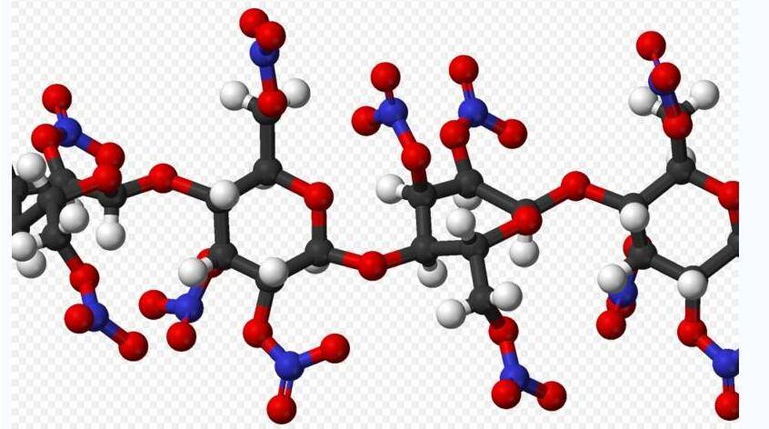
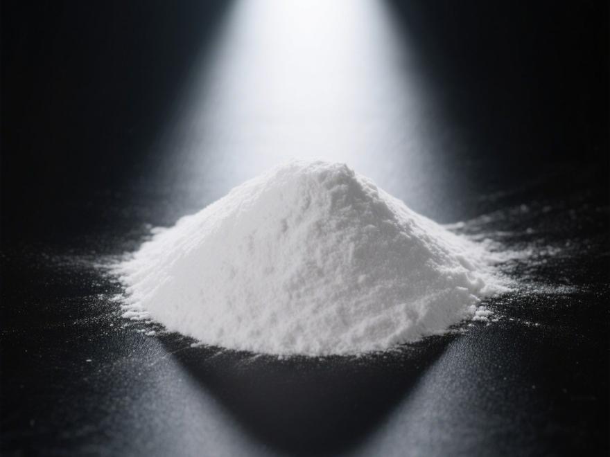
D mannose has the chemical formula C6H12O6 (structural formula shown in Figure 1). At room temperature and standard pressure, it exists as a white crystalline powder with a sweet taste and a slight bitter aftertaste, and has a low caloric value. D-mannose is widely distributed in nature: it is an important component of carbohydrates in body fluids and tissues, such as serum globulins, egg mucoproteins, and polysaccharides containing D-mannose in cell surface receptors [1]; the cell walls of plants such as palm seeds and coconut shells contain polysaccharides composed of D-mannose, and free D-mannose is also present in some fruits such as citrus fruits, peaches, and apples [2]. Currently, D-mannose is primarily produced through plant extraction, chemical synthesis, and bioconversion processes. With the deepening of research on D-mannose and the expansion of its application areas, market demand is continuously growing.


D-mannose in the human body plays a crucial role in maintaining normal cellular communication functions, including cell-to-cell communication, adhesion, signal transmission, and reception. It also plays an important role in the human immune defense system. Deficiency or overexpression of D-mannose may increase susceptibility to and severity of various diseases [3–5]. In recent years, the physiological functions of D-mannose have become a research hotspot. D-mannose intervention has been found to play an important role in preventing and alleviating urinary tract infections and antitumor effects [6–7], and may serve as an important functional raw material and bioreactor modifier in functional foods and biomedical fields. This review summarizes the current research progress on the production processes, metabolic pathways, and physiological functions of D-mannose.


D Mannose Powder 99%

1 Production processes of D-mannose

Currently, the main methods for producing D-mannose include plant extraction, chemical synthesis, and bioconversion. In the past, commercially available D-mannose was extracted from plants or produced chemically using D-glucose as a raw material. However, these methods are limited by raw material supply, energy consumption, by-products, or complex purification steps in downstream processes. Therefore, biological methods for producing D-mannose have garnered increasing attention in recent years. The research progress of different production processes is summarized in Table 1.


1.1 Plant extraction method

The plant extraction method involves hydrolyzing (acid hydrolysis, enzymatic hydrolysis, etc.), filtering, separating, purifying, and concentrating polysaccharides and oligosaccharides from plants and fruits to extract D-mannose. Palm kernel, coffee grounds, Brazilian berry seeds, and jujube powder are good raw materials for producing D-mannose [8–9]. In the literature, the temperature for plant extraction methods generally ranges from 50 to 121 °C, with the reaction time controlled between 30 minutes and 4 hours [10–12]. The specific extraction conditions and yields for D-mannose are shown in Table 1. Additionally, many microbial cell walls contain D-mannose polysaccharides, such as Saccharomyces cerevisiae and Pichia pastoris, which can also be used for D-mannose extraction [13]. The advantages of this method include a purely plant-based source, low cost, and high yield, making it suitable for industrial production. However, the production process requires high temperatures and high concentrations of acid/alkali solvents, which can cause environmental pollution, and production is significantly influenced by regional and seasonal factors.


1.2 Chemical synthesis method

The chemical synthesis method primarily uses molybdate catalysis to convert D-glucose into D-mannose through an isomerization reaction. Chemical synthesis requires strict control of reaction conditions, as shown in Table 1, with a temperature range of 100–150 °C, pH is around 3.0, and the reaction time is between 1 and 2 hours, with a conversion rate of D-glucose to D-mannose ranging from 29.2% to 44.8%. The advantage of the chemical synthesis method is the stable availability of raw materials, but the disadvantage is the high cost of raw materials. Additionally, due to the poor specificity of inorganic catalysts toward the substrate, the process often produces numerous byproducts, making separation difficult and increasing costs. Therefore, the chemical synthesis method still faces numerous challenges in production processes and is currently unsuitable for the industrial production of D-mannose.


1.3 Bioconversion Method

Bioconversion methods use D-fructose or D-glucose as raw materials and convert them into D-mannose through enzymatic reactions. Relevant enzyme genes can be identified from microorganisms, and recombinant expression plasmid vectors can be constructed to obtain recombinant engineered bacteria capable of producing high yields of the relevant enzymes, which are then used in enzymatic reactions to produce D-mannose [14]. The most studied enzyme is D-mannose isomerase. Additionally, D-lyxose isomerase can also be used to produce D-mannose. It is an aldose-ketose isomerase with broad substrate specificity, capable of catalyzing the isomerization reactions between D-xylose and D-lyxose, as well as between D-fructose and D-mannose [15]. Enzyme genes related to D-mannose production have been successfully isolated from bacteria such as Pseudomonas, Streptomyces, and Escherichia coli, with Escherichia coli commonly used as a host for engineering [14, 16].


Enzymatic production of D-mannose has been studied extensively, with production conditions and yields reported in other literature, as summarized in Table 1. During production, enzyme activity is significantly influenced by temperature and pH. As shown in Table 1, the optimal temperature range is 45–60°C, the pH range is 6.5–9.0, the reaction time is 1–8 hours, and the conversion rate ranges from 22.1% to 39.3%. The advantages of enzyme-mediated production of D-mannose include stable raw material sources, mild reaction conditions, and low costs. However, the isomerase enzymes used in current reports exhibit low catalytic efficiency and are easily influenced by reaction conditions.


2 Metabolic pathways of D-mannose

Ingested D-mannose is absorbed and metabolized in the intestine. The first step of the metabolic process involves the use of hexokinase (HK) and adenosine triphosphate (ATP) as cofactors to convert D-mannose into mannose-6-phosphate (M-6-P) through phosphorylation. The second step can be divided into three metabolic pathways: first, M-6-P is converted into D-fructose-6-phosphate (F-6-P) by mannose-6-phosphate isomerase (PMI); which then enters the glycolytic pathway; second, it is converted into 2-keto-3-deoxy-D-glycero-D-galactonoate-9-phosphate (KDN-9-P) by 2-keto-3-deoxy-D-glycero-D-galactonoate-9-phosphate synthase (KPS), further synthesizing KDN; third, M-6-P can be converted into mannose-1-phosphate (M-1-P) by phosphomannose isomerase (PMM), M-1-P reacts with guanosine triphosphate (GTP) via guanosine diphosphate mannose pyrophosphorylase (GMPP) to produce guanosine diphosphate mannose (GDP-M) and pyrophosphate, GDP-M serves as a D-mannose donor and can be biosynthesized into D-mannose phosphate (Dol-P-M) by D-mannose phosphate polyol synthase (DPM). These intermediates then participate in glycosylation reactions, attaching D-mannose to glycoproteins and glycolipids, including N-glycosylation, O-mannosylation, C-mannosylation, and glycosylated phospholipid-anchored protein synthesis. In addition to exogenously ingested D-mannose, glucose and glycogen can be converted into glucose-6-phosphate (G-6-P), which is then converted into F-6-P by phosphoglucose isomerase (PGI). F-6-P and M-6-P can be interconverted via phosphomutase (PMI).  A simplified diagram of the metabolic pathway of D-mannose is shown in Figure 2 [26–27].


Studies have shown that the catabolic metabolism of D-mannose is almost identical to that of glucose. In mammalian cells, 95%–98% of D-mannose enters the cell and is catabolized via PMI, with approximately 2% used for glycosylation. The utilization of M-6-P is largely dependent on the ratio of PMI to phosphomannose isomerase within the cell; a higher ratio promotes catabolism, while a lower ratio favors the glycosylation pathway [28–29]. Additionally, F-6-P derived from glucose can also be converted into D-mannose via PMI, followed by further metabolic processes.


3 Physiological functions and applications of D-mannose

In recent years, with the deepening of research on D-mannose, its various physiological functions have been discovered, including prevention and alleviation of urinary tract infections, antitumor effects, and immune system regulation. D-mannose can prevent pathogenic Escherichia coli in the urinary tract from adhering to mannose-containing glycoproteins, thereby exerting a protective effect on the urinary system. Second, ingested D-mannose can accumulate in tumor cells in the form of M-6-P, inhibiting enzymes related to glucose metabolism, or by reducing the expression of glucose transport carrier proteins, thereby interfering with glucose uptake by tumor cells and exerting antitumor effects; third, it has been found that D-mannose can enhance the conversion of initial T cells into regulatory T cells (Treg cells), thereby regulating the immune system. Additionally, D-mannose has been found to possess certain skin care benefits. Therefore, it is predicted that D-mannose will find increasingly widespread applications in the fields of health foods and biomedicine, with significant market potential.


3.1 Prevention and Alleviation of Urinary Tract Infections

Urinary tract infections are inflammatory conditions caused by bacterial invasion of the urinary tract, with Escherichia coli being the most common urinary tract pathogen [30]. E. coli possesses specific virulence factors, the most common being type I pili adhesion protein FimH, which helps bacteria adhere to D-mannose-containing glycoproteins on urinary epithelial cells, triggering an inflammatory response. This interaction is considered a signaling cascade [31–32], with the specific mechanism illustrated in Figure 3.


When D-mannose is excreted through the urethra, it can bind to the adhesion protein FimH at the tip of type I pili, saturating it and preventing the induction of the signaling cascade, thereby inhibiting bacterial adhesion to the urinary tract epithelium. Studies have demonstrated that D-mannose has excellent inhibitory effects on urinary tract infections.  LENGE et al. [33] conducted a systematic review and meta-analysis and found that D-mannose, administered at doses ranging from 420 mg to 2 g, with frequencies of 1 to 3 times daily for 1 week per month, demonstrated good tolerability and minimal side effects, with only a small proportion of participants experiencing diarrhea. It also demonstrated protective effects against recurrent urinary tract infections (compared to placebo), with efficacy comparable to antibiotics. KYRIAKIDES et al. [34] analyzed eight studies on D-mannose and found that intervention with D-mannose at doses of 500 mg to 2 g over different time periods (3 days to 6 months) reduced the incidence of recurrent urinary tract infections, prolonged the interval between recurrent infections, and improved patients' quality of life. Compared with natural D-mannitol, chemically modified D-mannitol exhibits higher affinity for FimH, and this D-mannitol glycoside molecule can bind more tightly to bacteria through pili [35–37].


Additionally, studies have shown that D-mannose and cranberry can enhance the therapeutic efficacy of antibiotics. RDULESCU et al. [38] conducted a preliminary randomized study involving 93 healthy non-pregnant women with uncomplicated urinary tract infections, with an average age of 39.77 ± 10.36 years, who were treated with either antibiotics alone or in combination with cranberry extract and D-mannose. The results showed that adding cranberry extract and D-mannose to antibiotics resulted in a higher cure rate at day 7 (84.4% vs 91.6%), and a significantly improved cure rate for drug-resistant strains (37.5% vs 88.8%). These results indicate that cranberry extract and D-mannitol, when used in combination with antibiotics, have a higher cure rate for urinary tract infections and enhance the sensitivity of urinary pathogens to antimicrobial therapy for acute urinary tract infections.


The above research data indicate that D-mannose has relatively clear preventive and alleviating effects on urinary tract infections, with good tolerability and high safety. Additionally, it has been found that combining D-mannose with other substances that have urinary system protective effects can produce better results. Overseas, there are already products containing D-mannose or D-mannose combined with cranberry extract and vitamin C, marketed for maintaining urinary system health, such as NOW Foods' D-Mannose Capsules, Clinicians' Mannose & Cranberry Urinary Tract Support Powder, and GNC's Cranberry + Mannose Extract. However, D-mannose has not yet been approved in China as a food ingredient or food additive, so it cannot currently be used in food products.


3.2 Anti-tumor

3.2.1 D-Mannose Interferes with Glucose Transport and Metabolism in Tumor Cells

Normal human cells and tissues primarily rely on aerobic oxidation for energy. However, tumor cells undergo changes in sugar metabolism. Under oxygen-rich conditions, malignant tumor cells exhibit abnormal glycolytic activity, primarily relying on glycolysis for energy, characterized by high glucose uptake rates, and high levels of metabolic byproduct lactic acid [39]. GONZALEZ et al. [40] found that compared to galactose, fructose, and glucose, D-mannose more effectively inhibits tumor cell proliferation. The possible mechanism is that D-mannose is phosphorylated via HK to form M-6-P, which accumulates in tumor cells in the form of M-6-P, M-6-P inhibits three enzymes involved in glucose metabolism: hexokinase, phosphoglucose isomerase, and glucose-6-phosphate dehydrogenase, thereby disrupting further sugar metabolism and inhibiting tumor cell proliferation.


Additionally, Wang Hao et al. [41] used colorectal cancer cell lines HCT116 and HT29 in vitro and primary colorectal tumor mouse models to find that D-mannose may interfere with glucose uptake and cell proliferation in colorectal cancer cells by reducing the expression of the glucose transporter protein GLUT1, thereby lowering the tumor incidence and progression in primary colorectal tumor mouse models. This suggests that D-mannose may inhibit tumor cell growth and proliferation by interfering with glucose transport and metabolism, thereby achieving an antitumor effect.


Current research literature demonstrates that D-mannose exerts certain effects on diseases such as lung cancer, liver cancer, colorectal cancer, and pancreatic tumors. Ge Hong et al. [42] found that 11.1 mmol/L D-mannose can inhibit non-small cell lung cancer cell lines A549 and H460, significantly increase the radiosensitivity and apoptosis rate of H460 cells, and exhibit more pronounced inhibitory effects on H460 cells as D-mannose concentration increases. Shan Tingting [43] found that D-mannose intervention could inhibit the proliferation and migration of human liver cancer cell line HepG2. Gonzalez et al. [40] further found that compared with the use of antitumor drugs alone, D-mannose combined with cisplatin or doxorubicin (antitumor drugs) could increase tumor cell apoptosis, and similar effects were also observed in mouse models, reducing tumor tissue in mice and improving tumor mouse survival rates. They also found that the sensitivity of different tumor cells to D-mannose depends on the level of phospho-D-mannose isomerase (PMI), and when PMI levels are low, tumor cells are more sensitive to D-mannose. Consistent with these findings, Yi Junyu et al. [7] also found that D-mannose not only inhibits the proliferation of breast cancer cells but also enhances their sensitivity to doxorubicin (a cancer treatment drug), and breast cancer cells with lower PMI expression are more sensitive to D-mannose.


3.2.2 D-mannose-modified anticancer drug carriers

D-mannose can also exert antitumor functions by modifying anticancer drugs. Compared with normal cells, malignant tumors overexpress D-mannose receptors, and D-mannose can be used for selective targeting and delivery of drugs [44]. By using D-mannose-modified nanodelivery systems to specifically deliver cytotoxic drugs to tumor cells, cancer cells can be directly induced to undergo apoptosis, thereby reducing toxicity to normal cells.


Some studies have shown that D-mannose-modified surfaces of nanocarrier systems enhance drug targeting to tumor cells and improve antitumor efficacy. Commonly used nanocarrier systems include polymer micelles, liposomes, and nanoparticles [45]. Wang Shijang [46]  conjugated D-mannose with a copolymer of poly(lactic acid-hydroxyethyl acid) and polyethylene glycol, and then bound it to the outer surface of gefitinib nano-mixed micelles, which promoted drug accumulation in lung cancer A549 cell tissue and induced apoptosis. Additionally, using a nude mouse lung cancer xenograft model, it was found to slow tumor growth rate and reduce tumor tissue proliferation ratio.


Chen Jing [47] modified glycyrrhizic acid liposomes with D-mannose-modified cholesterol ligands to prepare liver-targeted drugs. Using HepG2 cells as a cellular model, it was found to increase glycyrrhizic acid uptake in HepG2 cells, enhancing cell proliferation inhibition and promoting apoptosis. Furthermore, through drug intervention (dose of 5.25 mg/mL), the drug demonstrated liver-targeting properties in blood drug concentration tests in New Zealand rabbits and tissue distribution experiments in Kunming mice. Additionally, studies have shown that D-mannose combined with anticancer drug nanoparticles can also enhance therapeutic effects. By linking D-mannose with methotrexate-free nanoparticles via hydrolyzable ester bonds, drug release can be achieved through dual induction by lysosomal acidity and esterases, enhancing specific recognition, reducing drug dosage, and lowering toxicity to normal cells and tissues. In vitro (human breast cancer cell line MCF-7) and in vivo (MCF-7 human breast cancer nude mouse [BALB/c] model) experiments demonstrated that D-mannose enhances drug antitumor activity, exhibiting good therapeutic efficacy and biosafety [48]. However, before clinical application, extensive experimental data must be obtained, and its safety in humans must be verified [49].


Additionally, photodynamic therapy is one of the anticancer therapies. To reduce the damage of photodynamic therapy to normal cells, D-mannose can be used to modify photosensitizers, enhancing their selectivity toward cancer cells, thereby representing a novel, safe, and efficient targeted photodynamic therapy [50]. Cai Ying et al. [51] prepared D-mannose-modified photosensitizer nanoparticles using β-cyclodextrin and adamantane supramolecular recognition, which exhibited uniform particle size and good stability in solution. These nanoparticles were specifically recognized and taken up by breast cancer cells MDA-MB-231 overexpressing D-mannose receptors, and exhibited targeted photodynamic therapy effects on MDAMB-231 cells under 665 nm LED light irradiation.


In summary, D-mannose can inhibit glucose metabolism-related enzymes in tumor cells through the accumulation of M-6-P, thereby interfering with further glucose metabolism, or reduce glucose uptake by tumor cells by lowering the expression of glucose transport carrier proteins, thereby exerting antitumor effects. On the other hand, due to the overexpression of D-mannose receptors in tumor cells, D-mannose-modified anticancer drugs can enhance targeting, strengthen the inhibitory effect on tumor cells, and reduce toxicity to normal cells. Therefore, D-mannose holds great potential in tumor therapy.


3.3    Immune Regulation

3.3.1   Effects of D-mannose intervention on type 1 diabetes and airway inflammation

Some studies have found that D-mannose exerts immune regulatory effects by enhancing the conversion of initial T cells into regulatory T cells (Treg cells). Treg cells are essential immune regulatory cells that play a crucial role in inducing and maintaining immune tolerance, as well as preventing and suppressing autoimmune diseases [52]. ZHANG et al. [53] used non-obese diabetic mice with Treg cell deficiency as a model of type 1 diabetes and found that D-mannose can prevent and suppress type 1 autoimmune diabetes. It also prevents and suppresses the development of pulmonary airway inflammation in a mouse model of egg protein-induced asthma, and increases the proportion of Foxp3+ regulatory T cells in mice. Further studies revealed that the mechanism involves D-mannose promoting the activation of TGF-β in its latent form, thereby inducing the differentiation of initial CD4+ T cells into Treg cells. The activation of TGF-β (one of the most important immune inhibitory cytokines) is mediated through upregulation of integrin αvβ8 and increased reactive oxygen species.


3.3.2 Effects of D-mannose intervention on systemic lupus erythematosus

Similarly, D-mannose can also improve symptoms of systemic lupus erythematosus by regulating immune cells such as Treg cells. WANG et al. [54] first found that D-mannose can inhibit the maturation of bone marrow-derived dendritic cells (BMDCs) and the proliferation and activation of antigen-specific CD4+ T cells induced by BMDCs, while increasing the circulating frequency of Foxp3+ regulatory T cells in normal C57BL/6 mice. Subsequently, using a graft-versus-host disease (cGVHD) lupus-like mouse model and a B6.lpr spontaneous lupus mouse model, it was found that D-mannose treatment reduced the production of autoantibodies while decreasing the frequency of effector memory and helper T cells, as well as germinal center B cells and plasma cells. These findings indicate that D-mannose improves autoimmune activation in lupus models, at least in part by increasing Treg cells and downregulating the induction of immature dendritic cells and effect T cell activation.


3.3.3 Effects of D-mannose intervention on osteoporosis

Studies have shown that exacerbated inflammation disrupts bone metabolism. T cells can inhibit bone formation mediated by bone marrow mesenchymal stem cells (BMMSCs) by regulating interferon IFN-γ and tumor necrosis factor TNF-α levels. Treg cells can suppress T cells, reduce the secretion of pro-inflammatory cytokines, thereby improving BMMSC-based cranial defect repair [55]. Therefore, D-mannose may reduce osteoporosis in mice by increasing Treg cell proliferation. LIU et al. [56] administered D-mannose in drinking water to 12-month-old aged C57BL6/J mice and 8-week-old ovariectomized C57BL6/J mice for two months, compared to the untreated group, the cortical bone volume and trabecular microstructure were significantly increased in the D-mannitol intervention group, while the levels of osteoclast-related cytokines in bone marrow were downregulated, and the number of Treg cells in the spleen was increased, indicating that D-mannitol may alleviate osteoporosis in mice caused by aging and estrogen deficiency by inducing the proliferation of regulatory T cells.


The above results suggest that D-mannose, as a monosaccharide beneficial to human health, may have broad applications in promoting immune tolerance and treating/preventing human diseases associated with autoimmune and allergic conditions in the future.


3.4 Skin Care

The dermal reticular layer of the skin contains abundant collagen fibers, elastic fibers, and reticular fibers, conferring the skin with significant elasticity and resilience. During skin aging, fibroblasts lose their ability to synthesize collagen, collagen degradation increases, and the skin's biomechanical properties decline.


Research has found that D-mannose may have the potential to improve skin biomechanical properties. MEUNIER et al. [57] found that a complex composed of D-mannose, M-6-P, sodium phosphate, glycerol, and water could reverse visible signs of aging by reorganizing the dermal collagen network and improving skin biomechanical properties. When applied to human skin tissue for 7 days, it can increase the expression of three proteins—type V collagen, aging key protein antigen, and health antigen protein—and increase the density of the reticular dermis. Additionally, compared to the placebo group, it significantly increases facial collagen density, reduces the number of crow's feet wrinkles, and decreases the volume and depth of neck wrinkles. However, literature on the skin care functions of D-mannose is relatively limited, and this function requires further extensive research for confirmation.


4 Summary and Outlook

In recent years, D-mannose has garnered increasing attention from the food and pharmaceutical industries. Continuous in-depth research into its production processes and biological characteristics has revealed that, compared to plant extraction methods and chemical conversion methods, the biological method for producing D-mannose offers significant advantages. Furthermore, D-mannose is a highly promising “signal sugar” with potential functional and economic value, It can exert multiple physiological functions through different pathways: first, it can prevent bacterial adhesion to the urinary tract epithelium to prevent urinary tract infections; second, it can inhibit cancer cell proliferation by interfering with glucose transport and metabolism, and also modify anticancer drug carriers to selectively target and deliver anticancer drugs; third, it can promote the increase of Treg cells to exhibit immune regulatory effects in autoimmune diseases.


Further research data on D-mannose is needed to support its application in functional foods and the biopharmaceutical industry. In terms of production processes, to improve D-mannose yield and reduce production costs, further research is needed on enhancing the catalytic efficiency of isomerase enzymes and optimizing process conditions for bioconversion methods. In terms of physiological functional applications, the mechanism by which D-mannose prevents and alleviates urinary tract infections is relatively well-established, and it has been added to dietary supplements primarily targeting urinary tract protection in women. However, research on D-mannose's anti-tumor, immune-modulating, and skin care functions is relatively new. A deeper understanding of its mechanisms of action, exploration of other potential pathways, and specific applications and advantages in different product categories require further research data to support these claims. Additionally, the long-term safety of D-mannose remains to be further confirmed. D-mannose holds promise for playing a more active and significant role in functional foods and clinical therapies.


Reference

[1]FENG D, SHI B, BI F, et al. Elevated serum mannose levels as a marker of polycystic ovary syndrome[J]. Frontiers in Endocrino- logy,2019,10:711.

[2]TENG  B  W.  Preparation  of mannose  and  its   functional health function[J]. Light Industrial Science and Technol-   ogy,2014(7):67−68.

[3[LIU D R. Study on the prognostic value of mannose receptor and mirNA-1260B ingastroin- testinal tumors[D]. Lanzhou: Lanzhou University, 2017.

[4]MEI X. Experimental study on the effect of sinomenine on man- nose receptor expression and related antibodies in rats with rheuma- toid arthritis[D]. Chengdu: Chengdu University of Traditional  Chi- nese Medicine, 2014.

[5]HUANG C R, LI T P, WANG J, et al. Expression   and clinical significance of serum soluble mannose receptor in  pa-   tients with hepatocellular carcinoma[J]. Journal of Anhui Medical   University,2021,56(7):1127−1131.

[6[LU G S, SONG   B, JIN X Y. Research progress on the defense mechanism of urinary   tract  infection[J].  Journal  of  Third  Military  Medical  University,2000,22( 11):1116−1118.

[7]YI J Y, WANG Y Y, LIU X P, et al. Effect of mannose   on the proliferation of breast cancer cells and chemotherapeutic sensitivityof epirubicin[J]. Journal of Qingdong University: Med,2020,57(3):350−355.

[8[MUSSATTO S I, CARNEIRO L M, SILVA J P A, et al. A study on chemical constituents and sugars extraction from spent co- ffee grounds[J]. Carbohydrate Polymers,2011,83(2):368−374.

[9[WANG B. Chemical characterization and Ameliorating effect of polysaccharide from Chinese jujube on intestine oxidative injury by ischemia and reperfusion[J]. International Journal of Biological Macromolecules,2011,48(3):386−391.

[10]TAO Z, PAN Z, CHAO Q, et al. Isolation and purification of D-mannose from palm kernel[J]. Carbohydrate research,2009,344 ( 13):1687−1689.

[11]PEI J, SHENG Y H, GAO L Q. Preparation of high purity D-mannose[P]. China: CN105087712A, 2015-11-25.

[12]MONTEIRO A F, MIGUEZ I S, SILVA J P R B, et al. High concentration and yield production of mannose from aaí (Euterpe ol- eracea Mart.) seeds via mannanase-catalyzed hydrolysis[J]. Scien- tific Reports,2019,9( 1):10939.

[13[HU C H, YIN Y, YUE Q. Research progress  of mannoprotein  in  Saccharomyces  cerevisiae[J].  Wine Making,2007,34(4):64−66.

[14]WU H, ZHANG W, MU W. Recent studies on the biological production of D-mannose [J]. Applied Microbiology  and  Biotech- nology,2019,103(21-22):8753−8761.

[15[HUANG J, CHEN Z, ZHANG W, et al. D-lyxose isomerase and its application for functional sugar production[J]. Applied Mi- crobiology & Biotechnology,2018,102(6):2051−2062.

[16[ZHANG J, TONG Q Y. Optimization of the fermentation medium for produc- ing D-mannose-isomerase from Pseudomonas No. 2120[J]. Biopro-cess,2010( 1):37−42.

[17[ZHAO G H, WANG G B, PING L J, et al. The orthogonal experiment analysis on the producing mannose from dextrose by cat- alyzing effect of ammonium molybdate[J]. Shenyang Chemical In- dustry,2005( 1):39−41.

[18[HU H, LIU S, ZHANG W, et al. Efficient epimerization of glucose  to  mannose  over  molybdenum-based  catalyst  in  aqueous media[J]. Chemistry Select,2020,5(5):1728−1733.

[19]JIN P, DU QZ, WANG Y Y. A novel D-mannose-isomerase and its application[P]. Zhejiang: CN 111944796A, 2020-11-17.

[20[HUA X H, LI Y X, MA J W, et al. Characteri-   zation of D-mannose isomerase from Streptomyces and its  applica-   tion in the preparation of D-mannose [J]. Chinese Journal of Micro-   biology,2021,48(6):1930−1941.

[21]YU L N. Characterization of D- lysol isomerase and enzymatic preparation of mannose[D]. Wuxi: Jiangnan University, 2017.

[22[HUA X, LI Y, JIANG Z, et al. Biochemical properties of a   novel d-mannose isomerase from Pseudomonas syringae for D-mannose production[J]. Applied Biochemistry and Biotechnology,2021,193(5):1482−1495.

[23]HU X, ZHANG P, MIAO M, et al. Development of a recom- binant  d-mannose isomerase  and  its  characterizations  for D-Man- nose synthesis[J]. International Journal of Biological Macromole- cules,2016:328−335.

[24[LIU Z, LI Y, WU J, et al. A Novel Pseudomonas geniculata AGE family epimerase/isomeraseand its application in D-mannose synthesis[J]. Foods,2020,9( 12):1809.

[25[ZHANG W, HUANG J, JIA M, et al. Characterization of a novel D-lyxose isomerase  from  Thermoflavimicrobium  dichotomi- cum and its application for D-mannose production[J]. Process Bio- chemistry,2019,83(8):131−136.

[26]HU X, SHI Y, ZHANG P, et al. D-mannose: Properties, pro- duction, and applications: An overview[J]. Comprehensive Reviews in Food Science and Food Safety,2016,15(4):773−785.

[27[SHARMA  V,  ICHIKAWA  M,  FREEZE  H  H.  Mannose metabolism: More than meets the  eye[J].  Biochem  Biophys  Res Commun,2014,453(2):220−228.

[28[GORDON A, MARTIN H, RALF N, et al. Direct utilization of mannose for mammalian glycoprotein biosynthesis[J]. Glycobio- logy,1998(3):285.

[29[SHARMA V, FREEZE H H. Mannose efflux from the cells a potential  source  of  mannose  in  blood[J].  Journal  of  Biological Chemistry,2011:286.

[30]YANG J, XIONG J X, XIONG W W. Investigation and analysis of pathogen distribution and drug resistance of urinary tract infection[J]. Lab Med,2012,30(5):125−126.

[31[WANG J F. Effect offimH gene on the adhesion function of urinary tract pathogenic Escherichia coli type 1 pili [D]. Tianjin: Tianjin Medical University, 2014.

[32[HATTON N E, BAUMANN C G, FASCIONE M A. Developments   in   mannose-based    treatments   for   uropathogenic   Es-   cherichia coli induced urinary tract infections[J]. Chem Bio Chem,2020,22(4):613−629.

[33[LENGE R  S, BRADLEY M S, THOMAS D, et al. 39: D-mannose vs other agents for recurrent urinary tract infection preven-   tion  in  adult  women:  A  systematic  review  and  meta-analysis[J].   American  Journal   of  Obstetrics   and   Gynecology,2020,222(3): S797−S798.

[34]KYRIAKIDES R, JONES P, SOMANI B K. Role of D-man- nose in the prevention of recurrent urinary tract infections: Evidence from a systematic review of the literature[J]. European Urology Fo- cus,2020,7(5):1166−1169.

[35[JARVIS C,  HAN  Z,  KALAS  V,  et  al.  Antivirulence   isoquinolone  Mannosides:  Optimization  of  the  biaryl   aglycone  for FimH lectin binding affinity and efficacy in the treatment of chronic UTI[J]. Chem Med Chem,2016,11(4):367−373.

[36[HAN Z, PINKNER J  S, FORD B, et al. Lead optimization studies on FimH antagonists: discovery of potent and orally bioavail- able ortho-substituted biphenyl mannosides[J]. Journal of  Medici- nal Chemistry,2012,55(8):3945−3959.

[37]SPAULDING CN, KLEIN R D, RUER S, et al. Selective de- pletion of uropathogenic E. coli from the gut by a FimH antagonist [J]. Nature,2017,546(7659):528−532.

[38[RDULESCU, DAVID, TURCU, et al. Combination of cranberry extract and D-mannose-possible enhancer of uropathogen sensitivity to antibiotics in acute therapy of urinary tract infections: Results of a pilot study[J]. Experimental and Therapeutic Medicine,2020,20(4):3399−3406.

[39[LIU G, SONG G B. The role of glucose metabolism in tumor therapy[J]. Journal of Biomed- ical Engineering,2019(4):691−695.

[40[GONZALEZ P S, O'PREY J, CARDACI S, et al. Mannose impairs tumour growth and enhances chemotherapy[J]. Nature, 2018, 563(7733).

[41]WANG H, LI H T. Mannose inhibits the occurrence and proliferation of  col- orectal cancer by down-regulating GLUT1[J]. Food and Fermenta- tion Industry,2020,406( 10):58−64.

[42[GE H, LUO H, LIU K D, et al. Effect of mannose on radiosensitivity of 6 human non-small cell lung cancer cell lines[J].   Chin J Radiol Oncology,2020,29(7):558−562.

[43]SHAN T T. Inhibitory effect of mannose on human hepatoma cell line HepG2[D]. Changsha:  Hu- nan University of Traditional Chinese Medicine, 2020.

[44]CHU X X, BU F X, YIN T J, et al. Antitumor strategies based on targeted regulation  of tumor-associated  macrophages[J].  Journal  of China Pharmaceutical University,2021,52(3):9.

[45[YANG M, GAO Y T, ZHANG X Y, et al. Saccharide modified nanoscale delivery system[J]. Chem- istry of Life,2021,41(3):10.

[46[WANG S J. Development and antitumor  study of mannose-bound ph-sensitive targeted delivery of gefitinib nanocarriers[D]. Jinan: Shandong Uni- versity, 2018.

[47[CHEN J. Liver target- ing study of glycyrrhetinic acid liposomes modified by mannosylat- ed  cholesterol  ligands[D].  Guangzhou:  Guangzhou  University  of Traditional Chinese Medicine, 2018.

[48[FAN Z, WANG Y, XIANG S, et al. Dual-self-recognizing, stimulus-responsive and  carrier-free  methotrexate-mannose   conju- gate nanoparticles with highly synergistic chemotherapeutic effects [J]. Journal of Materials Chemistry B,2020,8(9):1922−1934.

[49]WEI Z, HUANG L, CUI L, et al. Mannose: Good player and   assister in pharmacotherapy[J]. Biomedicine & Pharmacotherapy,2020,129:110420.

[50]YANG G L, QIN S, LI W J. Research progress in the application of novel nanomaterials in tumor photodynamic thera- py[J]. Shandong Medicine,2020,60(35):84−87.

[51]CAI Y, WANG S T, WEI P, et al. Prepara-   tion of d-mannose-modified micelles and its application in targeted   photodynamic  therapy[J].   Journal  of  Food  And   Biotechnology,2019,38( 10):43−51.

[52[YIN C X, HOU Z J. Research progress of regulatory T cells and   their cytokines in autoimmune diseases[J]. Medical Review,2021,27(7):1284−1290.

[53]ZHANG D, CHIA C, JIAO X, et al. D-mannose induces reg- ulatory T cells and suppresses immunopathology[J]. Nature Medi- cine,2017,23(9):1036.

[54]WANG H, TENG X, ABBOUD G, et al. D-mannose amelio- rates autoimmune phenotypes in mouse models of lupus[J]. Bmc Immunology,2021,22( 1):1−12.

[55]LIU Y, WANG L, KIKUIRI T, et al. Mesenchymal stem cell- based tissue regeneration is governed by recipient T lymphocytes via IFN-γ and TNF-α[J]. Nature Medicine,2012,17( 12):1594−1601.    [56]LIU H, GU R, ZHU Y, et al. D-mannose attenuates bone loss in mice via Treg cell proliferation and gut microbiota-dependent anti-inflammatory effects[J]. Therapeutic Advances  in Chronic  Disease,2020,11:1−17.

[57[MEUNIER M, CHAPUIS E, LAPIERRE L, et al. Mannose- hosphate complex and improvement in biomechanical properties of the skin[J]. Journal of Cosmetic Dermatology,2021,20(2):1−13.

Follow Us
Back to the List
Prev

Clinical Application Study on D Mannose and Diabetes

Next

D Mannose: What Is It Made From?

Need More Information, Please Get in Touch.