

 English
 French
 Spanish
 Russian
 Korean
 Japanese
Green Spring Technology's Hyaluronic Acid Drive Innovation in Medical Dressing Products
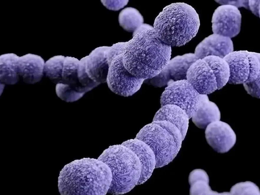
Hyaluronic acid is a natural polysaccharide composed of alternating D-glucuronic acid and N-acetylglucosamine units. It is widely distributed in the extracellular matrix, skin, and synovial fluid of living organisms, exhibiting exceptional hydrophilicity, antioxidant properties, rheological characteristics, and viscoelasticity.
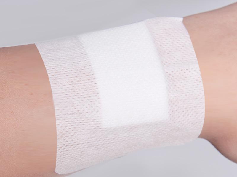
Green Spring Technology leverages advanced biopreparation techniques to supply hyaluronic acid powder across multiple molecular weights, suitable for developing premium wound care dressings. High-molecular-weight hyaluronic acid offers superior water retention and lubricating properties, aiding in maintaining a moist environment; low-molecular-weight variants are more readily absorbed and utilised, supporting skin repair processes.
Through Green Spring Technology's stable, high-quality hyaluronic acid raw materials solution, clients can develop more functional and comfortable dressing products, enhancing product differentiation and competitiveness to provide consumers with gentler, more effective care experiences.

1 Hyaluronic Acid: Exceptional Properties Empowering Premium Dressing Innovation
As a member of the glycosaminoglycan family, hyaluronic acid exhibits distinctive physicochemical properties due to its unique structure—unsulphated and not covalently bound to core proteins. It possesses outstanding hydrophilicity, rheological properties, and viscoelasticity, coupled with excellent antioxidant capacity, making it an ideal multifunctional matrix material.
Currently, various dressing products developed based on hyaluronic acid are gaining increasing market attention. Their outstanding moisturising properties and biocompatibility provide extensive scope for product design, enabling broad application across diverse personal care and professional repair scenarios.
Leveraging advanced biotechnology, Green Spring Technology is committed to supplying hyaluronic acid powder of varying molecular weights, high purity, and superior quality. This supports clients in developing innovative dressing products that better meet market demands, jointly driving industry technological advancement and product iteration.
1.1 Hydrophilicity
As a vital component of the extracellular matrix, hyaluronic acid possesses abundant hydroxyl and carboxyl groups within its molecular structure, conferring exceptional hydrophilic properties. This characteristic enables it to carry substantial negative charge, effectively attracting and binding water molecules and cations, thereby demonstrating outstanding moisture absorption and retention capabilities.
Owing to its potent water-binding capacity, hyaluronic acid is acclaimed as a ‘natural moisturising factor,’ finding extensive application in eye lubricants and skin hydration products. It delivers a gentle yet highly effective moisturizing solution for the personal care sector.
1.2 Fluidity and Lubrication Properties
As a biopolymer naturally present in synovial fluid, hyaluronic acid powder exhibits outstanding rheological characteristics and lubricating properties. It effectively reduces friction and alleviates pressure arising from mechanical movement.
In applications involving medical device interventions and human contact, hyaluronic acid is extensively researched as a mild, safe lubricant due to its excellent biocompatibility and high lubricity. Compared to traditional lubricants, hyaluronic acid exhibits higher purity and reduced allergenic potential, delivering a more comfortable user experience.
Currently, lubricant products developed from hyaluronic acid are progressively being applied across multiple professional care domains, offering new possibilities for advancing safer, more skin-friendly personal health solutions.
1.3 Viscoelasticity
At ambient temperatures, hyaluronic acid presents as a white, odourless solid powder, soluble in water but insoluble in organic solvents. Its aqueous solutions exhibit outstanding viscoelasticity and distinctive rheological behaviour, displaying typical non-Newtonian fluid characteristics.
Hyaluronic acid readily undergoes molecular modification to form structures of varying molecular weights. High-molecular-weight hyaluronic acid creates solutions with pronounced viscoelasticity, making it suitable for simulating the lubricating environment within biological systems. This provides an ideal material foundation for developing diverse care and functional products.
2 Properties of Hyaluronic Acid and Molecular Weight Correlation
Research indicates that the functional properties of hyaluronic acid (HA) are closely related to its molecular weight. Based on molecular weight (MW), hyaluronic acid can be categorised into several groups, each possessing distinct physicochemical characteristics and application potential:
· Hyaluronic acid oligosaccharides (O-HA, MW < 1×10⁴ Da): Characterised by low molecular weight, they are readily absorbed and participate in multiple biological processes;
· Low molecular weight hyaluronic acid (LMW-HA, 1×10⁴ Da < MW < 25×10⁴ Da): Exhibits excellent permeability and biological activity, suitable for products requiring high absorption rates;
· Medium molecular weight hyaluronic acid (MMW-HA, 25×10⁴ Da < MW < 100×10⁴ Da): Combines moisturising, lubricating and carrier functions, widely utilised in diverse skincare formulations;
· High molecular weight hyaluronic acid (HMW-HA, MW ≥ 1×10⁶ Da): Exhibits superior hydration, lubrication and viscoelastic properties, aiding protective barrier formation;
· Very high molecular weight hyaluronic acid (vHMW-HA, MW > 6×10⁶ Da): Possesses exceptional viscoelasticity and lubricating properties, suitable for demanding application environments.
Leveraging advanced production processes, Green Spring Technology provides a full spectrum of high-purity hyaluronic acid raw materials across all molecular weight ranges. This supports development needs across diverse application scenarios, empowering clients to create more innovative and safer personal and professional care products.
3 Hyaluronic Acid Enables Innovation in Multi-Form High-End Dressings
Hyaluronic acid, with its unique physicochemical properties—including exceptional hydrophilicity, viscoelasticity, and biocompatibility—has become an ideal raw material for developing high-end dressings in diverse forms. Currently, hyaluronic acid-based material technologies have been successfully applied to electrospun fibres, composite membranes, hydrogels, and sponges, delivering richer and more efficient solutions for personal and professional care markets.
Green Spring Technology is committed to supplying high-quality, multi-molecular-weight hyaluronic acid powder, supporting clients in the development and application of innovative dressing products. Together, we advance the evolution of care products towards functionalisation, comfort enhancement, and diversification.
3.1 Hyaluronic Acid-Based Electrospinning: Innovative Materials Empowering Premium Dressings
Electrospinning technology enables the production of ultra-fine fibres ranging from micrometre to nanometre scale, forming advanced materials with high porosity, high extensibility, and the capacity to load functional components.
Research indicates that combining hyaluronic acid with natural materials such as chitosan and gelatin can construct coaxial nanofibres with excellent biocompatibility.To address stability issues during spinning, auxiliary components like polyvinyl alcohol (PVA) and cyclodextrin are incorporated, effectively enhancing fibre formation and structural integrity. Further incorporation of nanofillers such as cellulose nanocrystals (CNCs) can also improve the fibres' adsorption capacity and mechanical properties.
Green Spring Technology supplies high-quality hyaluronic acid raw materials, supporting the innovative application of electrospinning technology in the high-end dressing sector and assisting clients in developing more competitive products.
3.2 Hyaluronic Acid-Based Films: Flexible Innovation, Empowering Diverse Applications
Hyaluronic acid-based film materials combine softness with elasticity, enabling functional optimisation through composite formulations with multiple components. Research demonstrates that composites of hyaluronic acid with proteins, polysaccharides, and nanomaterials yield films exhibiting high transparency, superior moisture retention, and excellent mechanical properties, suitable for applications demanding stringent material performance.
Green Spring Technology offers diverse modified hyaluronic acid raw materials, supporting the development of high-performance membrane materials and providing innovative foundational materials for the care and skincare industries.
3.3 Hyaluronic Acid-Based Hydrogels: High-Water-Content Materials Driving Technological Breakthroughs
Hyaluronic acid-based hydrogels represent a class of functional materials characterised by high water content and soft elasticity, demonstrating vast potential across multiple high-tech fields.
Research indicates that combining hyaluronic acid with functional components such as dopamine enables the construction of hydrogel systems with antioxidant properties, effectively enhancing material stability and environmental adaptability. Additional techniques employ physical electrostatic cross-linking and freeze-drying processes to develop modified hyaluronic acid hydrogels with outstanding performance, offering novel approaches to material design.
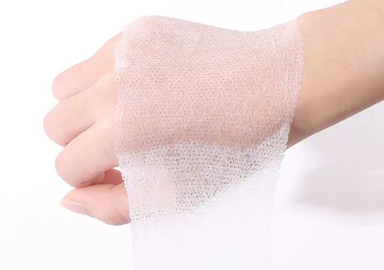
3.4 Hyaluronic Acid-Based Sponges: Porous Materials Driving Product Innovation
Hyaluronic acid-based sponge materials, characterised by high porosity, excellent breathability, and superior water absorption, represent an ideal choice for diverse high-end applications.
Research indicates that cross-linking with natural polymers such as chitosan and alginate significantly enhances the mechanical strength and structural stability of hyaluronic acid sponges. These porous materials feature a soft texture and high pore connectivity, delivering outstanding performance in moisture retention and gas exchange. They are particularly suited for scenarios demanding superior material properties and skin-friendly characteristics.
Green Spring Technology is dedicated to supplying high-quality hyaluronic acid raw materials, supporting the development and application of porous sponge materials, and providing innovative material solutions for personal care, functional skincare, and other sectors.
4 Outlook
Hyaluronic acid, with its exceptional physicochemical properties and biocompatibility, has become a key ingredient in premium care and professional health products.
As a specialised hyaluronic acid supplier, Green Spring Technology offers a comprehensive range of high-purity products across various molecular weights. These are suitable for developing premium dressings including electrospun fibres, membrane materials, hydrogels, and sponges. We are committed to delivering stable, reliable raw material solutions that enhance product performance and competitive differentiation.
Partner with Green Spring Technology to seize new opportunities in hyaluronic acid innovation. Contact us at helen@greenspringbio.com or WhatsApp: +86 13649243917 for further product and technical information.
Reference
[1]GRUPPUSO M,TURCO G,MARSICKh E,et al.Polymeric wound dressings, an insight into polysaccharide-based electrospun membranes[J].Applied Materials Today,2021,24:101148.
[2]DOVEDYTIS M,LIU Z J,BARTLETT S.Hyaluronic acid and its biomedical applications:A review[J].Engineered Regeneration,2020, 1:102-113.
[3]MARIANA F P G,SÓNIA P M,CÁTIA S D C,et al.Hyaluronic acid- Based wound dressings:Areview[J].Carbohyd Polym,2020,241:1-17.
[4]QIU Y B,MA Y Q,HUANG Y Y,et al.Current advances in the biosynthesis of hyaluronic acid with variable molecular weights[J]. Carbohydrate Polymers,2021,269:118320.
[5]SU S,BEDIR T,KALKANDELEN C,et al.Coaxial and emulsion electrospinning of extracted hyaluronic acid and keratin based nanofibers for wound healing applications[J].European Polymer Journal,2021,142:110158.
[6]SUNJ,PERRY S L,SCHIFFMAN J D.Electrospinning Nanofibers fromChitosan/HyaluronicAcidComplexCoacervates[J].Biomacromo lecules,2019,20(11):4191-4198.
[7]ABBAS Z B,ESMAEIL M,MILAD F,et al.Dual spinneret electrospun nanofibrous/gel structure ofchitosan-gelatin/chitosan-hyaluronic acid asa wound dressing:In-vitro and in-vivo studies[J].International Journal of Biological Macromolecules,2020,162:359-373.
[8]MORGANE S L,ANNE C C,SÉVERINE V,et al.Electrospinning in water and in situ crosslinking of hyaluronic acid/cyclodextrin nanofibers:Towards wound dressing with controlled drug release[J]. Carbohydrate Polymers,2018,207:276-287.
[9]YASMEIN H,ESMAIL M E,ELBADAWYl A K,et al.Electrospun PVA/hyaluronic acid/L-arginine nanofibers for wound healing applications:Nanofibers optimization and in vitro bioevaluation[J].InternationalJournal of Biological Macromolecules,2020,164:667-676.
[10]YIN CJ,QI XJ,WU J,et al.Therapeutic contact lenses fabricated by hyaluronic acid and silver incorporated bovine serum albumin porous filmsforthetreatmentofalkali-burned cornealwound[J].International Journal of Biological Macromolecules,2021,184:713-720.
-
Prev
Hyaluronic Acid Drives Innovation in Cosmetic Formulations
-
Next
How Hyaluronic Acid Elevates Your Cosmetic Formulations